Vinno P Series
Simplify your work with a touch
Excellent image quality
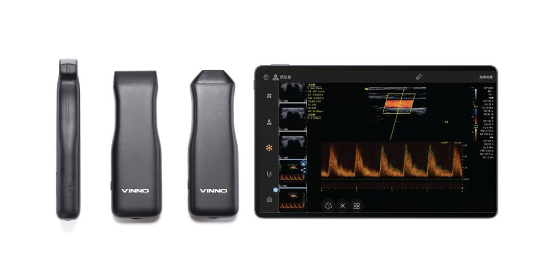

Standard Features
- CW/CF/PW/M
- wide range of probe frequencies (2–16MHZ)
- Advanced wireless data transfer
- Supports internal and external puncture guidance
- One click order image optimisation
- Biopsy guide and needle enhancement)

Design
- Touch operation
- Ergonomic, slimline design
- Convex, linear and phased array transducer
- Detachable lithium long life battery (unlimited reload–cycles)
- IPX7 waterproof protection

Technical Features
- Cloud storage
- Remote on-line software updates
- Connect to tablet or phone
- High-performance wireless data transfer, avoids data delay or loss
- Powerful data post processing capability

Good Image Quality
- SEE TABLE BELOW
IMAGE QUALITY INFORMATION
Obstetrics:
Good image quality allows for a more comprehensive fetal anatomy scan including
- Assessing fetal anatomy in greater detail, including evaluation of the brain, heart, spine, limbs, kidneys, and other organs to detect structural abnormalities.
- Assessment of amniotic fluid volume: Evaluating the amniotic fluid levels to assess for abnormalities such as polyhydramnios or oligohydramnios.
- Placental evaluation: Assessing the position, size, and structure of the placenta to detect placental abnormalities.
Abdominal:
Good image quality enables more detailed and accurate assessments of abdominal structures including
- Visualizing organ structures, such as the liver, kidneys, spleen, and pancreas, in greater detail.
- Measurements of organ dimensions.
- Assessment of organ texture.
- Identification of focal abnormalities, such as solid masses or simple fluid collections, can be performed more reliably.
Gynecology:
Detailed assessment of uterine structures including
- Visualizing and evaluating the endometrium, myometrium, and cervix for abnormalities like polyps, fibroids, or adenomyosis.
- Identification of adnexal masses: Detecting and characterizing complex ovarian masses, including solid or cystic tumors
Vascular:
With Good to Excellent image quality, vascular ultrasound allows for detailed assessment of arterial and venous structures throughout the body. It enables the evaluation of blood flow patterns, detection of stenosis or occlusion, identification of aneurysms, assessment of vascular grafts or fistulas, and guidance for vascular nterventions or surgeries.
It's important to note that achieving optimal image quality depends on various factors, including the ultrasound system's capabilities, the operator's skill and technique, patient factors (such as body habitus or patient cooperation), and the specific clinical indication for the ultrasound examination.