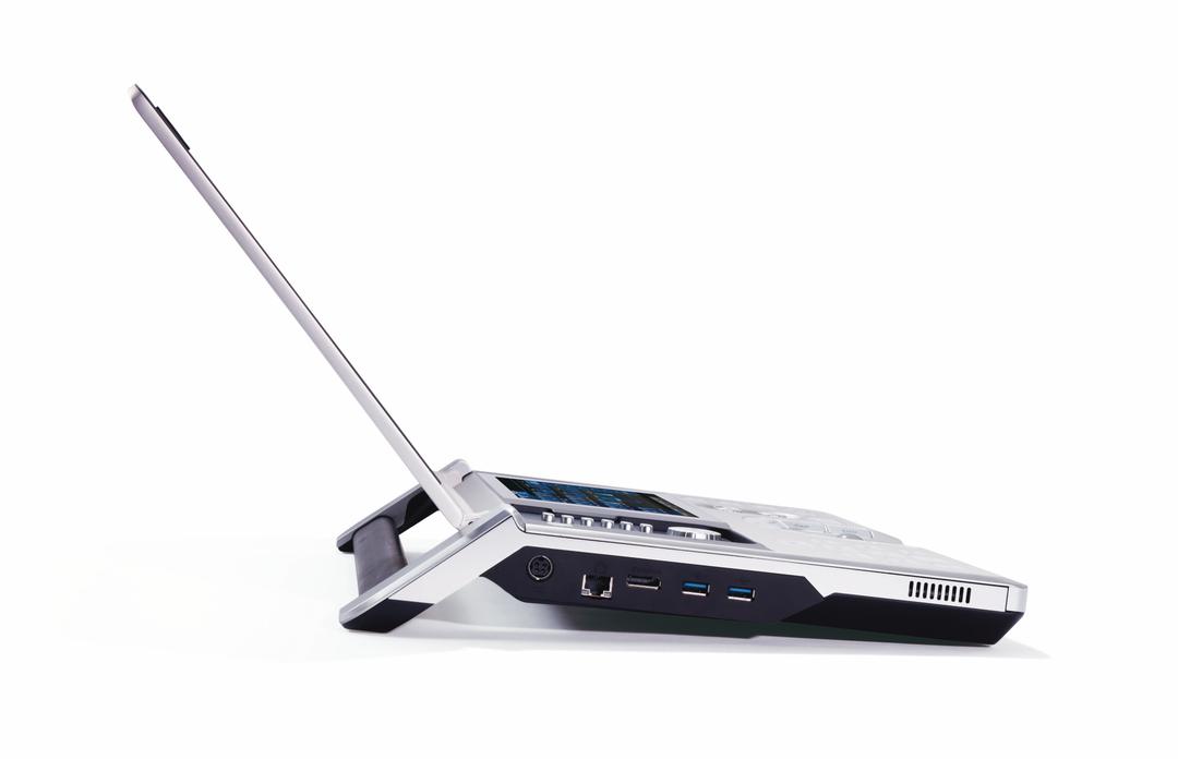
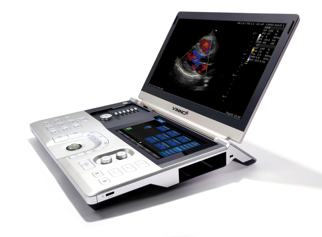
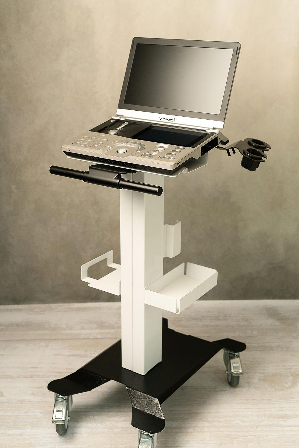
Vinno V6
Simplify your work with a touch
Excellent image quality


Standard Features
- CW/CF/PW/M
- Stress Echo
- Anatomical M Mode
- Auto EF
- Tissue Velocity Imaging
- Strain Imaging
- Elastography
- Customizable report system

Design
- Light Weight (3Kg)
- 15 inch high resolution screen
- 8 inch touch screen
- Standard Battery (80 Min scan time)
- Quick, intuitive work flow
- Multi probe port adapter

Technical Features
- 500Gb Hard drive
- Pacs
- Wi-Fi
- DICOM
- Cloud Storage
- Carry on Case

Good Image Quality
- SEE TABLE BELOW
IMAGE QUALITY INFORMATION
Obstetrics:
Advanced fetal neurosonography
- Detailed evaluation of the fetal brain, including assessment of specific brain structures, detection of subtle abnormalities, and identification of neural tube defects.
- Fetal echocardiography: Comprehensive assessment of fetal heart structure and function, including detailed evaluation of cardiac chambers, valves, and blood flow patterns.
- Assessment of fetal growth and well-being: Detailed measurements of fetal biometry, Doppler evaluations of blood flow in umbilical arteries, middle cerebral arteries, and evaluation of placental function.
Abdominal:
Excellent image quality provides the highest level of detail and clarity for abdominal scanning including
- Precise evaluations of intricate structures, such as liver segments, renal pyramids, or pancreatic ducts.
- Subtle abnormalities, such as small liver metastases, renal masses with specific features, or focal pancreatic lesions, can be detected.
- Advanced Doppler techniques, like evaluating portal vein flow or quantifying renal blood flow, can be performed accurately. Assessments for liver diseases, renal pathologies, pancreatic disorders, and complex abdominal surgeries.
Gynecology:
- Advanced evaluation of uterine abnormalities: Assessing complex uterine abnormalities such as congenital anomalies, uterine septum, or intrauterine adhesions.
- Advanced assessment of adnexal pathology: Characterizing complex adnexal masses in detail, including differentiating benign from malignant masses.
- Evaluation of deep infiltrating endometriosis: Visualizing and assessing the extent of endometriosis involvement in the pelvis, including evaluation of deep infiltrating lesions and their relationship to surrounding structures.