Vinno V5
Simplify your work with a touch
Excellent image quality
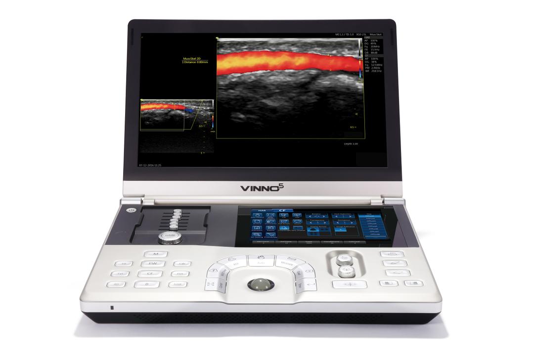



Standard Features
- CW/CF/PW/M
- High Quality 5D
- Auto Measurement
- Tissue Velocity Imaging
- Anatomical M Mode
- Elastography
- Auto EF

Design
- Ultra light compact design
- Intuitive touch screen
- Built in battery
- Multi port probe connector

Technical Features
- Technical Features
- Wi-Fi
- Blue Tooth
- Cloud Storage
- 500Gb hard Drive
- DICOM

Very Good Image Quality
- SEE TABLE BELOW
IMAGE QUALITY INFORMATION
Obstetrics:
Advanced fetal cardiac evaluation become possible including:
- Assessing fetal heart structure and function in more detail, including evaluation of cardiac chambers, valves, and blood flow patterns.
- Detailed evaluation of fetal brain structures: Visualizing and assessing the fetal brain anatomy, including the ventricles, cortex, and midline structures.
- Identification of soft markers: Detecting subtle signs that may indicate an increased risk for chromosomal abnormalities, such as nuchal translucency or echogenic intracardiac focus.
Abdominal:
Very good image quality allows for highly detailed evaluations of abdominal structures including:
- Detecting smaller or subtle abnormalities, such as small liver lesions, renal cysts with specific characteristics, or early signs of inflammation in organs
- Detailed assessments of organ vascularity, such as Doppler evaluations of blood flow in the liver or kidneys, can be performed accurately.
- Identification of small stones or sludge in the gallbladder or bile ducts becomes more feasible.
Gynecology:
Very good image quality allows for evaluation of endometrial pathology including:
- Assessing the endometrium in detail for abnormalities such as endometrial polyps, hyperplasia, or cancer
- Advanced assessment of adnexal pathology: Characterizing complex adnexal masses in detail, including differentiating benign from malignant masses.
- Evaluation of deep infiltrating endometriosis: Visualizing and assessing the extent of endometriosis involvement in the pelvis, including evaluation of deep infiltrating lesions and their relationship to surrounding structures.
Vascular:
With Good to Excellent image quality, vascular ultrasound allows for detailed assessment of arterial and venous structures throughout the body. It enables the evaluation of blood flow patterns, detection of stenosis or occlusion, identification of aneurysms, assessment of vascular grafts or fistulas, and guidance for vascular interventions or surgeries.
It's important to note that achieving optimal image quality depends on various factors, including the ultrasound system's capabilities, the operator's skill and technique, patient factors (such as body habitus or patient cooperation), and the specific clinical indication for the ultrasound examination.